- Peripheral lung nodules with nCLE
- Lesson 03/05
Malignant Nodule
Please note, nCLE is used in conjunction with transbronchial needle aspiration (TBNA) procedures to puncture and image lung nodules, and fits through a transbronchial needle with an inner diameter of 0.91 mm.
You may review the various image criteria and videos before doing some self training questions.

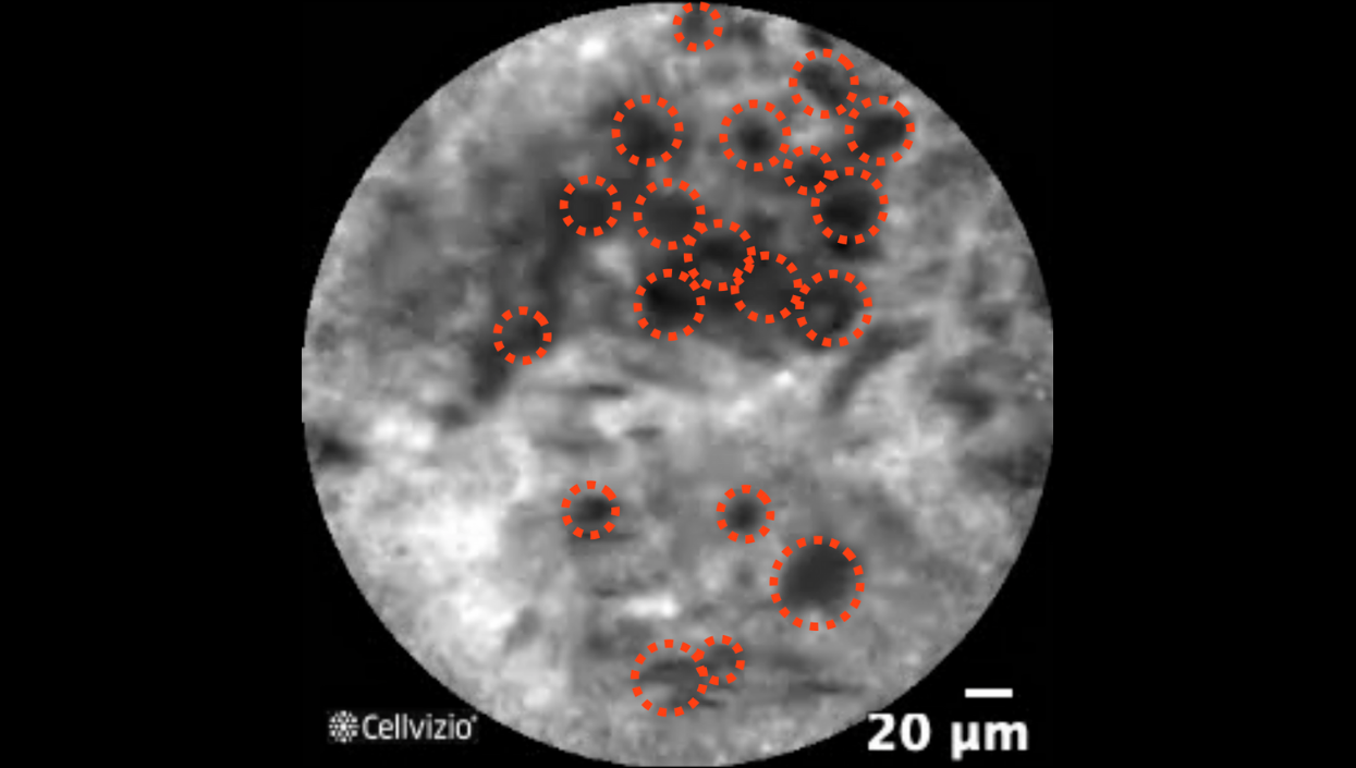
Enlarged pleomorphic cells
Organ: Lung
Investigated pathology: Peripheral lung nodule with nCLE - Malignant nodule
Probe type: AQ-Flex 19
Laser wavelength: 488 nm
Courtesy: Dr. Sandeep Bansal, DuBois Regional Medical Center, PA, USA

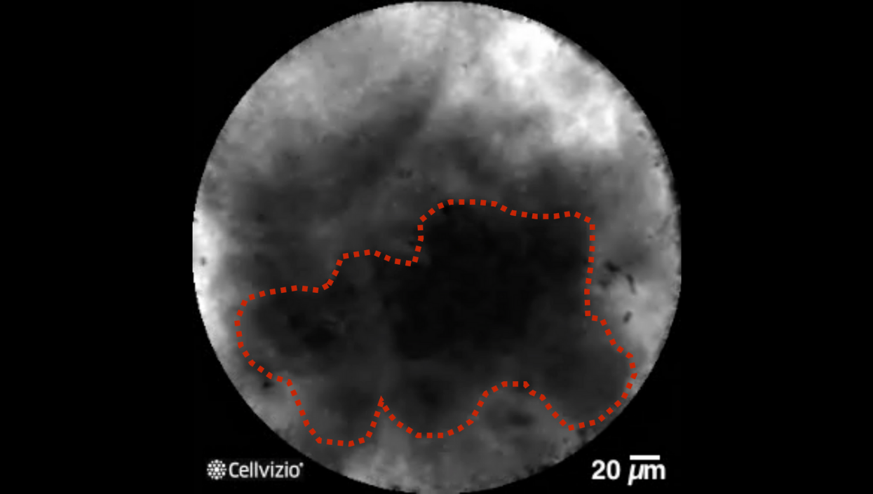
Dark clumps
Organ: Lung
Investigated pathology: Peripheral lung nodule with nCLE - Malignant nodule
Probe type: AQ-Flex 19
Laser wavelength: 488 nm
Courtesy: Prof. dr. Jouke T. Annema and Dr. Tess Kramer, Amsterdam UMC, the Netherlands

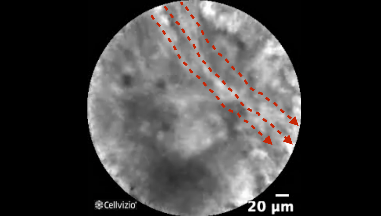
Directional streaming
Organ: Lung
Investigated pathology: Peripheral lung nodule with nCLE - Malignant nodule
Probe type: AQ-Flex 19
Laser wavelength: 488 nm
Courtesy: Dr. Sandeep Bansal, DuBois Regional Medical Center, PA, USA
PLAY THE TRAINING VIDEOS:
The following criteria are visible in the videos: enlarged pleomorphic cells, dark clumps, and directional streaming.
Organ: Lung
Investigated pathology: Peripheral lung nodule with nCLE - Malignant nodule
Probe type: AQ-Flex 19
Laser wavelength: 488 nm
Courtesy: Dr. Sandeep Bansal, DuBois Regional Medical Center, PA, USA
Organ: Lung
Investigated pathology: Peripheral lung nodule with nCLE - Malignant nodule
Probe type: AQ-Flex 19
Laser wavelength: 488 nm
Courtesy: Dr. Sandeep Bansal, DuBois Regional Medical Center, PA, USA
These criteria have been reported and assessed in clinical studies. Other criteria of interpretation might exist. Thus whenever possible, confirmatory evaluation via conventional histopathology or any other clinical information is recommended.
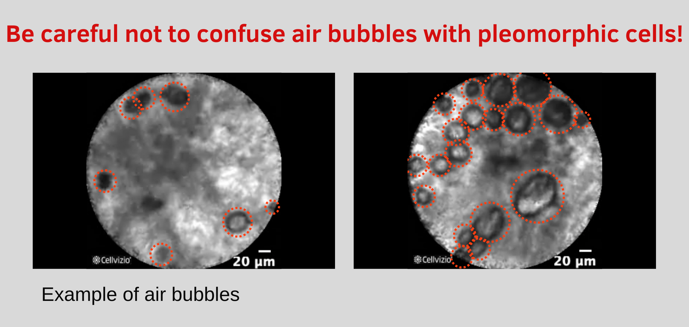
Air Bubbles

Tip: Sometimes very small air bubbles can look like malignant cells. As they can look similar, need to wait until you no longer see air bubbles before judging the video, or try to image another location.
AIR BUBBLE EXAMPLE VIDEOS:
Organ: Lung
Investigated pathology: Peripheral lung nodule with nCLE - Air bubbles
Probe type: AQ-Flex 19
Laser wavelength: 488 nm
Courtesy: Dr. Sandeep Bansal, DuBois Regional Medical Center, PA, USA
Organ: Lung
Investigated pathology: Peripheral lung nodule with nCLE - Air bubbles
Probe type: AQ-Flex 19
Laser wavelength: 488 nm
Courtesy: Dr. Sandeep Bansal, DuBois Regional Medical Center, PA, USA
