- Lung nodules with pCLE
- Lesson 01/03
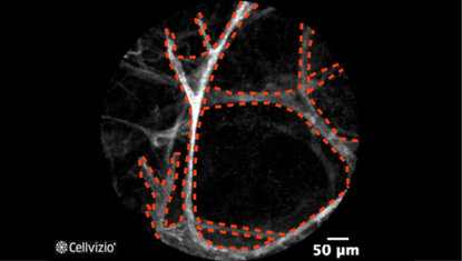
Healthy alveoli
Please note, pCLE is used in conjunction with standard bronchoscopic procedures with a working channel inner diameter of 1.9 mm. It can only image autofluorescent microstructures in the lung, such as the elastin fibers in the alveolar septi, vessel walls, and bronchial airways.
You may review the various image interpretation sets before doing some self training questions.

Thin regular alveolar septi

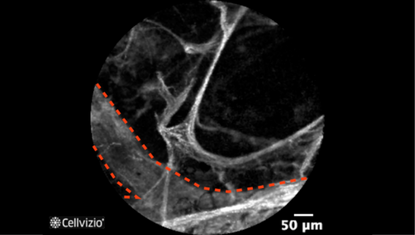
Blood vessel
PLAY THE TRAINING VIDEOS:
Organ: Lung
Investigated pathology: Peripheral lung nodule
Probe type: AlveoFlex
Laser wavelength: 488 nm
Courtesy: Dr. David Wilson, Columbus regional hospital, Columbus, IN, USA
Organ: Lung
Investigated pathology: Peripheral lung nodule
Probe type: AlveoFlex
Laser wavelength: 488 nm
Courtesy: Dr. Adam Wellikoff and Dr. Robert Holladay, Louisiana State University, Shreveport, LA, USA
These criteria have been reported and assessed in clinical studies. Other criteria of interpretation might exist. Thus whenever possible, confirmatory evaluation via conventional histopathology or any other clinical information is recommended.
