- Pancreatic Cysts during EUS-FNA
- Lesson 01/06
Normal Pancreas
You may review the various image interpretation sets before doing some self training questions.

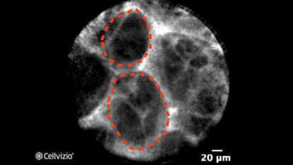
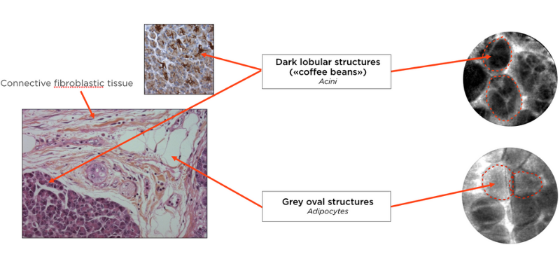
Acini: Dark lobular structures («coffee beans»)

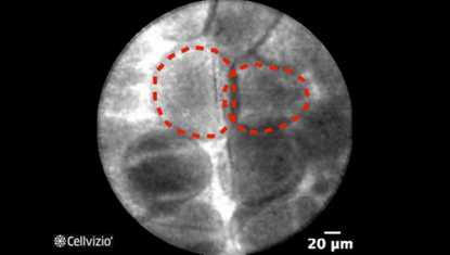
Adipocytes: Grey oval structures
PLAY THE TRAINING VIDEOS:
Organ: Pancreas
Investigated pathology: Pancreatic cyst
Probe type: AQ-FLEX 19
Laser wavelength: 488 nm
Courtesy: The CONTACT clinical trial, 2013
These criteria have been reported and assessed in clinical studies. Other criteria of interpretation might exist. Thus whenever possible, confirmatory evaluation via conventional histopathology or any other clinical information is recommended.
Pathology Correlation
The pancreas is typically composed of dark-staining cells in clusters called acini, of lightly stained clusters of cells called islets of Langerhans, and of fat cells called adipocytes.
True pancreatic cysts are typically composed of a wall separating the inside of the cyst from the pancreatic parenchyma. The epithelial structure of the wall including its cytology help determine the nature of the cyst, notably for serous cystadenomas, IPMN, and mucinous cystadenomas, which count among the main types of cysts. Pseudocysts, on the contrary, are lined with scar or inflammatory tissue.
The AQ-Flex™ 19 Confocal Miniprobe™ has a depth of imaging located between 40 and 70 microns, and is introduced inside the cysts through the wall. It allows imaging the inner wall opposite the puncture and therefore can identify the epithelial structures of the wall and help differentiate them.
What happens in the normal pancreas?
Correlation between normal pancreas and endomicroscopy images:
Konda et al. (1) showed that pancreatic endomicroscopy can identify histological elements in the pancreatic parenchyma. Endomicroscopy images are obtained when the probe is pushed either further the cystic wall or if too much pressure is exerted on the cystic wall.
Endomicroscopy revealed images of acini, which appear as dark lobular structures similar to «coffee beans». Adipocytes appear as grey oval structures, either round when seen separately or in a polygonal network when packed as clusters.

References:
1) Konda VJ, Aslanian HR, Wallace MB, Siddiqui UD, Hart J, Waxman I (2011) First assessment of needle-based confocal laser endomicroscopy during EUS-FNA procedures of the pancreas (with videos). Gastrointest Endosc 74:1049-60

