- Pancreatic Cysts during EUS-FNA
- Lesson 02/06
Serous cystadenoma
You may review the various image interpretation sets before doing some self training questions.

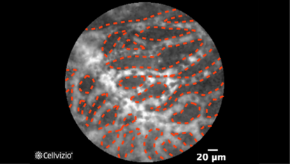
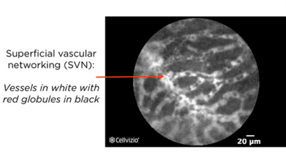
Superficial vascular networking
PLAY THE TRAINING VIDEOS:
Organ: Pancreas
Investigated pathology: Pancreatic cyst
Probe type: AQ-FLEX 19
Laser wavelength: 488 nm
Courtesy: The CONTACT clinical trial, 2013
Organ: Pancreas
Investigated pathology: Pancreatic cyst
Probe type: AQ-FLEX 19
Laser wavelength: 488 nm
Courtesy: The CONTACT clinical trial, 2013
These criteria have been reported and assessed in clinical studies. Other criteria of interpretation might exist. Thus whenever possible, confirmatory evaluation via conventional histopathology or any other clinical information is recommended.
Pathology Correlation
What happens in a serous cystadenoma?
Correlation between serous cystadenoma and endomicroscopy images:
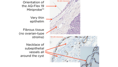
Napoléon et al. (2) showed that pancreatic endomicroscopy can identify specific structures underneath the cyst wall epithelium of a serous cystadenoma.
The presence of a necklace of subepithelial vessels all around the cyst has been shown by immunohistochemistry by using the anti-CD31 antibody vascular marker. These vessels are just below the epithelium (approx. 20 microns below) which explains that they can be observed with the AQ-Flex™ 19. They form a very dense network of thin vessels, and the real-time and dynamic imaging of Cellvizio enables to visualize the circulation of the blood cells within these vessels. It is called the Superficial Vascular Network (SVN). Moreover, a very thin epithelium (less than 20 microns thick) lines up the wall of serous cystadenomas, yet it is too thin to be visualized by confocal endomicroscopy.
Serous cystadenoma observed with an optical microscope:


This section has been prepared with the collaboration of Anne-Isabelle Lemaistre, MD.
References:
2) Napoleon B, Lemaistre AI, Pujol B, Caillol F, Mialhe-Morellon B, Giovannini M (2014) In vivo characterization of pancreatic cystic tumors by needle-based confocal laser endomicroscopy (nCLE). Proposition of a comprehensive classification. Gastrointest Endosc 79:AB434-35

