- Gastric Diseases
- Lesson 06/07
High Grade Intraepithelial Neoplasia
You may review the various image interpretation sets before doing some self training questions.
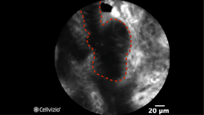
Prominent distorted pits (first example)
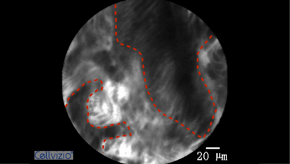
Prominent distorted pits (second example)
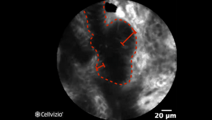
Variable width of the epithelial lining (first example)
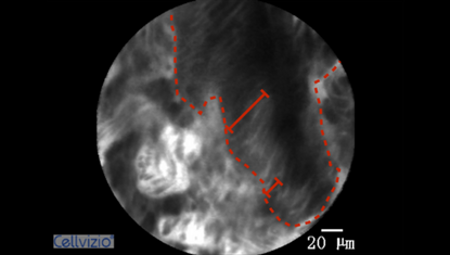
Variable width of the epithelial lining (second example)
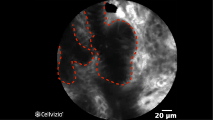
Bundled cells form a continuous bar (first example)
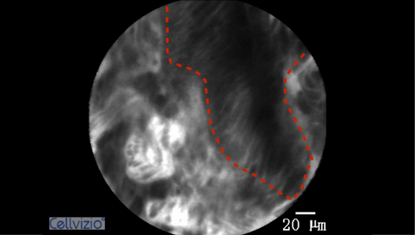
Bundled cells form a continuous bar (second example)
PLAY THE TRAINING VIDEOS:
Organ: Stomach
Investigated pathology: Gastric disease
Probe type: GastroFlex UHD
Laser wavelength: 488 nm
Courtesy: Dr. Zhen Li, Institute Shandong University, Qilu Hospital, China
Organ: Stomach
Investigated pathology: Gastric disease
Probe type: GastroFlex UHD
Laser wavelength: 488 nm
Courtesy: Dr. Zhen Li, Institute Shandong University, Qilu Hospital, China
These criteria have been reported and assessed in clinical studies. Other criteria of interpretation might exist. Thus whenever possible, confirmatory evaluation via conventional histopathology or any other clinical information is recommended.
