- Pancreatic Cysts during EUS-FNA
- Lesson 03/06
Intraductal Papillary Mucinous Neoplasm
You may review the various image interpretation sets before doing some self training questions.

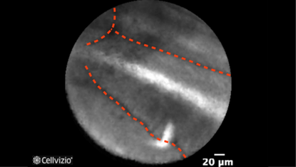
Papillary projection (first example)

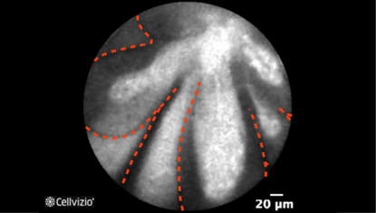
Papillary projection (second example)
PLAY THE TRAINING VIDEOS:
Organ: Pancreas
Investigated pathology: Pancreatic cyst
Probe type: AQ-FLEX 19
Laser wavelength: 488 nm
Courtesy: The INSPECT clinical trial, 2012
Organ: Pancreas
Investigated pathology: Pancreatic cyst
Probe type: AQ-FLEX 19
Laser wavelength: 488 nm
Courtesy: The CONTACT clinical trial, 2013
These criteria have been reported and assessed in clinical studies. Other criteria of interpretation might exist. Thus whenever possible, confirmatory evaluation via conventional histopathology or any other clinical information is recommended.
Pathology Correlation
What happens in an Intraductal Papillary Mucinous Neoplasm (IPMN)?
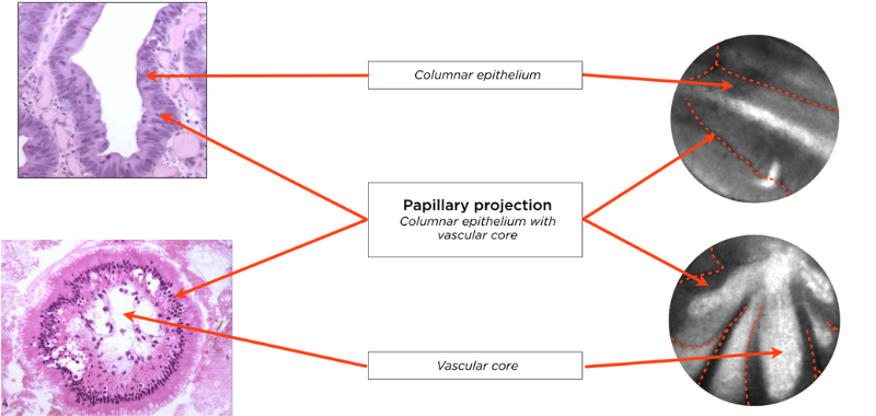
Correlation between an IPMN and endomicroscopy images:
Konda et al. (3) showed that pancreatic endomicroscopy can identify cytological elements in the wall of an IPMN.
The presence of papillae in IPMN can be observed with Confocal Endomicroscopy. The columnar epithelium as well as the vascular core, which composes a papillae, are recognizable. As such, on endomicroscopy images, a papillary projection is recognizable either as two dark borders (the epithelial borders) lining a white core (the vascular core), or as a dark ring (the columnar epithelium) around a white core (the vascular core).

This section has been prepared with the collaboration of Anne-Isabelle Lemaistre, MD.
References:
3) Konda VJ, Meining A, Jamil LH, Giovannini M, Hwang JH, Wallace MB, Chang KJ, Siddiqui UD, Hart J, Lo SK, Saunders MD, Aslanian HR, Wroblewski K, Waxman I (2013) A pilot study of in vivo identification of pancreatic cystic neoplasms with needle-based confocal laser endomicroscopy under endosonographic guidance. Endoscopy 45:1006-13

